Brain MRI: An Unclear Lesion — What Now?
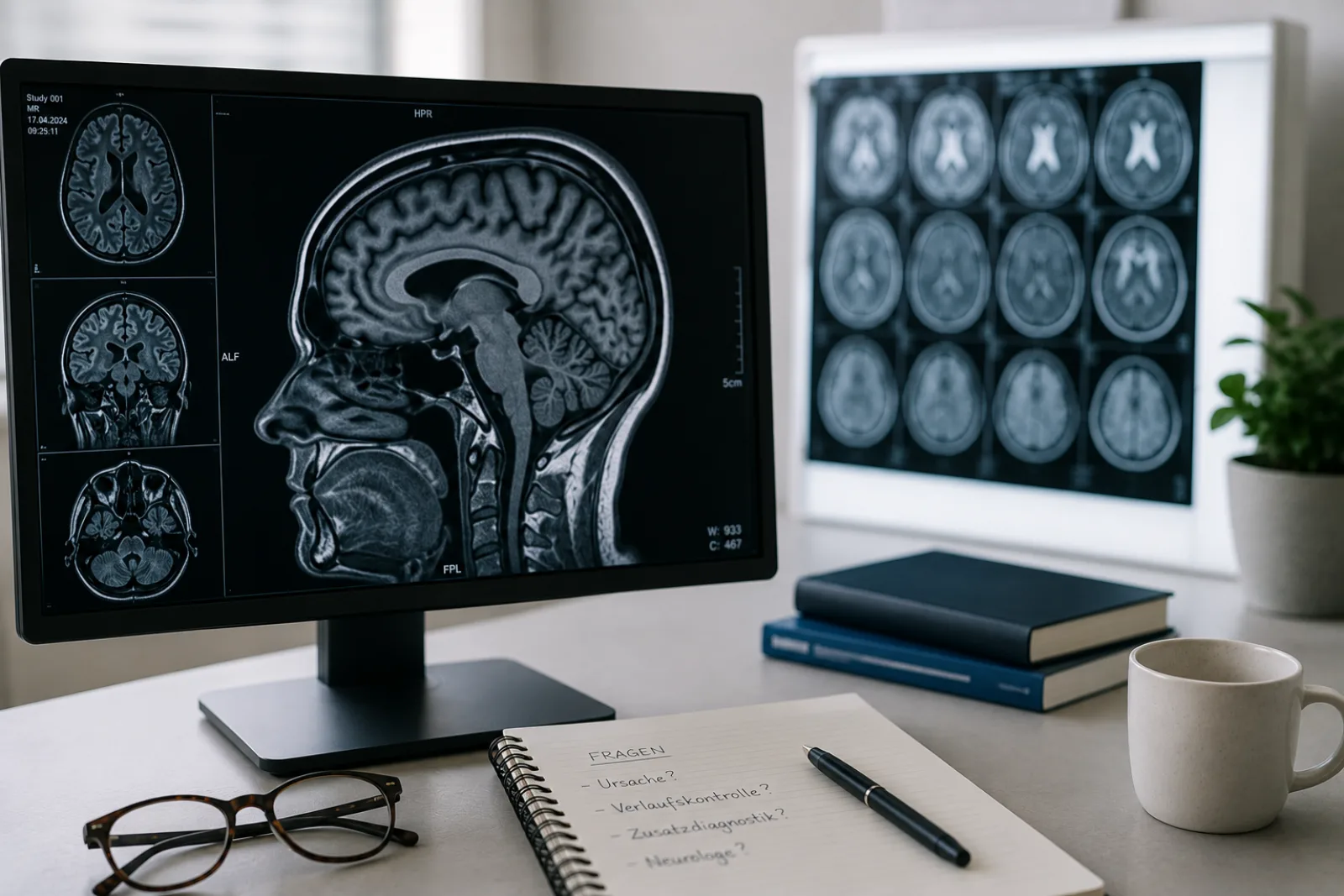
Two pages of MRI report. Sequences, slice thicknesses, anatomical landmarks — most of it you can skim. Then comes a sentence like this: “Scattered T2-hyperintense lesions in the white matter, etiology not clearly determinable.” And suddenly your eye stops. Not at the technical detail — at the one word you half understand: lesion.
Search for it and you land on multiple sclerosis and brain tumors within a minute. Both are possible, both are rare — and an evening of internet research makes neither more nor less likely. What helps is a calm look at what the report actually says.
This article puts the most common phrases from a brain MRI report into plain language. It explains why a radiologist writes “unclear,” what can lie behind brain lesions — and what the next sensible step is. It will not replace the conversation with your doctor, but it should leave you better equipped for it.
A second opinion on your report
Second View is a German service for radiological second opinions on MRI, CT, and X-ray. If your report has left you with questions, you can upload your images and documents here and have a specialist go through them independently. We review what you send, then quote you a price, usually €70–200. Your written second opinion normally arrives within 48 hours of payment.
What “lesion” actually means in the report
The word sounds like injury, like damage. In radiology it means something more sober: a change in tissue. On the MRI images, the radiologist sees a spot that differs from the brain tissue around it — in signal intensity, in shape, or at its margins. “Lesion” describes that difference. It says nothing about the cause and nothing about the severity.
This matters: “Lesion” is a descriptive word, not an alarm word. It says that something is visible — not that something bad is going on.
Some phrases that may show up in your report, and what they mean:
“T2-hyperintense foci in the white matter” — On certain MRI images (the T2 or FLAIR sequences), individual spots in the brain’s white matter look brighter than the tissue around them. These are the “white spots” patients keep running into in their own research.
“Focal signal increase of unclear etiology” — At one small, well-defined spot, the brain tissue shows an altered signal. The images alone cannot say reliably what caused it. “Etiology” means cause — and “unclear” means several causes are possible.
“Nonspecific white matter lesions” — Changes in the brain’s white matter that do not point to any one disease. The radiologist chooses “nonspecific” on purpose, because the images alone do not allow a definite classification.
When brain lesions are an emergency
Before this article works through the possible causes, one clear line: certain neurological symptoms call for immediate action — no matter what the MRI report says.
If one or more of the following symptoms comes on suddenly, go straight to the nearest emergency department or call emergency services (112, the European emergency number):
- Sudden paralysis or numbness in the face, arm, or leg — especially on one side
- Sudden trouble speaking or understanding speech
- Sudden loss or blurring of vision in one or both eyes
- A sudden, extremely severe headache with no obvious cause
- Sudden dizziness with an unsteady gait
- Clouded consciousness or sudden confusion
- A seizure, if it is your first
These symptoms can signal a stroke or another acute injury to the brain. The medical terms include ischemic stroke (a blockage of blood flow) and intracerebral hemorrhage. In this situation, every minute counts.
An “unclear lesion” on its own, with no acute neurological symptoms, is not an emergency. It is a reason to look carefully — not a reason to drive to the emergency department.
Not sure what your report is telling you
This is where experience counts. At Second View, brain scans go to radiologists who read neuroimaging regularly — the difference between someone who sees a handful of these a year and someone who sees them every week.
Why a radiologist writes “unclear” — and what that says about the finding
An MRI of the brain shows tissue differences in high contrast. It shows where something has changed and what the change looks like. What it does not show is why. Different processes — age-related vascular changes, inflammation, scarring after an infection, more rarely a tumor — can look alike on MRI.
When a radiologist writes “unclear” or “not clearly determinable,” they are doing exactly the right thing professionally: naming the range of possible diagnoses instead of committing to a cause the image alone cannot support. That is not helplessness; it is diagnostic care.
A typical phrase from a report shows how this works:
“DD demyelination vs. microangiopathic origin” — The radiologist names the two most likely causes: inflammatory damage to the insulating sheath around the nerve fibers (as happens in multiple sclerosis) or age-related vascular changes. Listing both means the MRI alone is not enough for a reliable classification. More information is needed — the clinical picture, your age, other conditions, and further testing if necessary.
The most common causes behind “unclear lesions”
Microangiopathic changes — wear on the small vessels
By far the most common cause of white spots on a brain MRI is microangiopathy: small tissue changes that follow the slow narrowing of the brain’s finest blood vessels. The risk factors are high blood pressure, diabetes, smoking, and age itself.
How common they are depends heavily on age, and the numbers are striking: at 50, about 5 percent of people have such changes; past 80, nearly everyone who gets scanned does [3]. A systematic review in the BMJ puts the prevalence at 11 to 21 percent among 64-year-olds and up to 94 percent among 82-year-olds [3].
Radiologists often grade how extensive these changes are on the Fazekas scale:
“Fazekas grade I” — A few scattered pinpoint bright spots. In most people over 50 this is a normal finding, of no clinical significance.
“Fazekas grade II” — The spots are starting to run together. That suggests a heavier vascular burden, often from long-standing high blood pressure.
“Fazekas grade III” — Extensive, merged changes reaching from the brain’s ventricles out into the surrounding white matter. At this point, treating the vascular risk factors consistently matters a great deal.
Fazekas grade I in someone over 60 is usually a finding of no consequence for health. Even so, it is worth keeping an eye on the vascular risk factors — blood pressure and blood sugar above all — because they influence how far the changes progress [3, 8].
Have a second radiologist look at your images
With small-vessel changes, what matters most is the trend over time. If you have older scans, upload them alongside the current one: comparing them is often what settles whether anything has actually changed.
Migraine-associated and post-infectious changes
Migraine can leave white spots on a brain MRI too. These lesions are generally small and pinpoint, do not take up contrast, and stay stable over time. As far as we know today, they are of no clinical significance in themselves — which reassures many patients, because at first sight the spots are easy to mistake for MS lesions.
The same goes for changes left behind by an infection: viral illnesses — including SARS-CoV-2 — can leave small spots in the white matter that stay visible on MRI without progressing or causing symptoms.
When multiple sclerosis is genuinely a possibility
Fear of MS is why so many patients panic after reading their report. Search the internet for “lesion in the brain” and you end up on MS information pages almost immediately. The reality is more nuanced.
MS lesions have a characteristic distribution: they cluster around the brain’s ventricles (periventricular), just beneath the cortex (juxtacortical), in the brainstem or cerebellum (infratentorial), and in the spinal cord. One telltale sign is what radiologists call “Dawson’s fingers” — elongated spots running perpendicular to the ventricles. The current McDonald criteria, reflected in the guideline of the German Society of Neurology — an S2k guideline, meaning one agreed on by a formal panel of experts — require more than the MRI picture for a diagnosis of MS: clinical symptoms, a spinal fluid analysis, and how things develop over time [5].
When MS-typical lesions turn up by chance — on an MRI ordered for headaches or dizziness, say — in someone with no MS symptoms, this is called a radiologically isolated syndrome (RIS) [6]. RIS is not a diagnosis of MS. Not everyone with a radiologically isolated syndrome goes on to develop MS. Conversion rates run at 13 percent within five years when no additional risk factors are present — and 34 percent when factors such as spinal cord lesions or positive spinal fluid findings are present [7]. Either way, a neurologist should be involved in the assessment.
An independent read of your scan
If MS is the worry, it is worth getting a clear answer rather than a search engine’s. A radiologist experienced in neuroimaging can tell you whether your images show the pattern MS produces, or one of the many other things that look similar at first glance.
Mass lesions — benign more often than you might think
Rare, but possible: the brain MRI shows a structure described as a mass. That sounds alarming — and yet the most common tumor inside the skull is a meningioma, a benign tumor of the membranes covering the brain. In more than 90 percent of cases it is WHO grade I and grows slowly. Many meningiomas are found by chance and never cause symptoms. In follow-up studies over two to five years, 50 to 70 percent show no tendency to grow at all.
Arachnoid cysts — fluid-filled pockets between the membranes around the brain — are another common incidental finding. They are usually present from birth, harmless, and need no treatment.
Lesions that take up contrast deserve particular attention. When a change on MRI brightens after contrast is given, that points to an active process — fresh inflammation, a tumor, or a vascular change. Findings like these always need further workup.
How the location of a lesion helps narrow things down
Where a lesion sits in the brain is a decisive piece of the puzzle for the radiologist. Some phrases you may find in the report:
“Periventricular” — around the brain’s ventricles. Both microangiopathic changes and MS-typical spots turn up here. Shape and distribution usually tell them apart.
“Subcortical” or “in the white matter” — in the white matter beneath the cortex. The range of possible causes is broad, from vascular to inflammatory to rare neurodegenerative diseases.
“Infratentorial” — beneath the tentorium, meaning in the region of the cerebellum and brainstem. Lesions here get extra attention, because they are less often purely vascular and more often point to inflammation or a mass.
The number matters too. A single, stable lesion is judged differently from several spots spread across different parts of the brain. Whether a lesion needs follow-up depends on its location, size, shape, signal behavior, and the clinical context — not on the fact that it is there.
What to do if your finding stays unclear
If your brain MRI report describes an unclear lesion, there are several sensible next steps.
1. Read the report calmly. The words “lesion” and “unclear” sound more threatening than they usually are. The explanations in this article can help you make sense of individual phrases.
2. Make an appointment with the doctor who ordered the scan. Go through the report together. Questions worth bringing along:
- Does this finding fit my age and my risk factors?
- Is a follow-up scan recommended — and if so, how soon?
- Would a referral to a neurologist or neuroradiologist make sense?
3. Follow-up imaging is the most useful tool you have. Staying unchanged over time is the surest sign that something is harmless. If a lesion looks the same on a follow-up MRI after six or twelve months, the odds rise considerably that it means nothing for your health.
4. Get your vascular risk factors under control. If the report mentions microangiopathic changes, have your blood pressure, blood sugar, and blood lipids checked. This does not treat the lesion; it keeps it from progressing.
5. Get a radiological second opinion. With an unclear finding, a second specialist assessment of the MRI images can bring clarity — often without repeating the scan. A radiologist with neuroradiology experience may recognize patterns the first reading could not pin down. This is no criticism of the first reader: differences between two radiological assessments happen and are part of the normal variability of medical diagnosis. The second opinion complements the first assessment with an additional specialist perspective.
How to get a second opinion
Before you agree to a biopsy, an operation, or a year of follow-up scans, it can be worth having the images read again. A second opinion from Second View gives you an independent assessment to weigh alongside the first — usually within 48 hours.
Frequently asked questions
Can brain lesions disappear again? Yes, some can. Fresh inflammatory spots — during an MS relapse, for instance — can shrink under treatment or fade over time, and changes left by an infection can recede as well. Microangiopathic changes, by contrast, generally stay put or slowly progress; they do not go away.
Do I need to see a neurologist now? Not in every case. A neurology consultation makes sense if an inflammatory cause is suspected, in younger patients with unclear white matter lesions, or if you have neurological symptoms. With clearly microangiopathic changes in later life, care from your primary care doctor is often enough — with an eye on the vascular risk factors.
How often should follow-up scans be done? That depends on the finding. For a newly discovered, unclear lesion, many guidelines recommend a follow-up MRI after six to twelve months. If it proves stable, the intervals can stretch out. Lesions that take up contrast, or spots that are growing, need closer follow-up.
Is a second opinion worthwhile if my radiologist called the finding “unclear” themselves? Especially then. “Unclear” means the imaging alone does not allow a reliable classification. A second look — ideally by a radiologist who specializes in neuroradiology — can sharpen the differential diagnosis, and often narrows the list of open possibilities. That is not a correction but an addition — and a standard part of careful diagnosis.
At Second View, an independent specialist reads your MRI images and puts the findings in context — one more perspective to take into the conversation with your doctor.
This article is for general information and does not replace an individual medical exam or consultation. If you have acute symptoms, neurological deficits, or any of the emergency signs described above, contact a doctor or an emergency department immediately.
Last medical review: May 11, 2026.
Questions about your MRI report
Getting started is straightforward: upload the images and paperwork you already have, and an independent radiologist reviews them. You see the price before anything is billed — usually €70–200 — and the written report follows within about 48 hours.