Endometriosis on MRI: What It Shows and When a Second Opinion Is Worthwhile
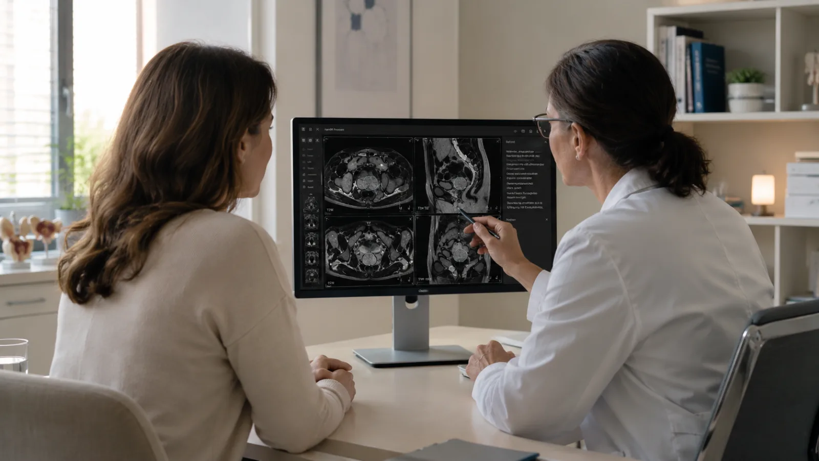
For many women, years go by between the first symptoms and a diagnosis of endometriosis. Crushing period pain, pain during sex or bowel movements, month after month of trying to get pregnant without success — and, in between, a string of appointments where nobody finds anything. Eventually someone orders an MRI of the pelvis, often to work things up before a possible operation.
What comes back usually reads one of two ways. Either a run of unfamiliar terms — endometrioma, deep infiltrating endometriosis, adenomyosis. Or one flat sentence, “no evidence of endometriosis,” while the pain goes on shaping your daily life. Either way, the same question follows: how much can this image be trusted?
This article lays out what an MRI can show in endometriosis and what it cannot, translates the most common terms in the report, and explains when a second radiological read is worth arranging. It is not an individual diagnosis or a treatment recommendation — those belong to the conversation with your own doctor.
The role of MRI in diagnosing endometriosis
In endometriosis, tissue resembling the lining of the uterus grows outside the uterine cavity — on the ovaries, on the peritoneum, between the vagina and the rectum, or elsewhere in the pelvis. It is a chronic condition, and how easily its lesions can be seen varies a great deal.
The first imaging test is not MRI but transvaginal ultrasound. Germany's national endometriosis guideline (an S2k guideline, meaning the medical societies formally agreed on it) calls ultrasound the standard first-line method. MRI comes second, as a way to fill in what ultrasound alone cannot settle.
ESUR, the European radiology society for urogenital imaging, names four situations where an MRI makes sense: the ultrasound is inconclusive; the ultrasound looks normal but the symptoms continue; an operation is being planned; or symptoms persist after surgery.
MRI does not replace ultrasound. It picks up where ultrasound runs out. What it does well is show deeper lesions and how far they reach, which is exactly what surgeons need before a procedure.
What MRI shows — and what it does not
MRI shows three forms of endometriosis reliably: cysts on the ovaries, deep-growing lesions, and endometriosis inside the wall of the uterus.
An ovarian cyst reads something like this in the report:
“Endometrioma of the left ovary, T1-hyperintense with T2 shading.”
In plain language: there is a cyst on one ovary filled with old blood — a “chocolate cyst,” as it is often called. The thickened blood gives off a distinctive signal on MRI, which makes these cysts fairly easy to identify.
Deeper lesions are described like this:
“Deep infiltrating endometriosis with a T2-hypointense nodule in the rectovaginal septum.”
In plain language: endometriosis tissue has grown past the surface of the peritoneum, in this case into the layer between the vagina and the rectum. On MRI it looks like a dark, scar-like nodule. Deep infiltrating lesions often sit on the supporting ligaments of the uterus, on the bladder, or on the bowel — places ultrasound has trouble reaching.
Endometriosis in the muscle of the uterus, adenomyosis, is recognized partly by a measurement:
“Junctional zone widened to 14 mm.”
In plain language: the inner muscle layer of the uterine wall is thickened. Once it passes about 12 millimeters, that points toward adenomyosis.
One form, though, routinely slips past imaging: superficial peritoneal endometriosis. These flat lesions are usually too small and too thin to register on MRI, and that is the method's biggest blind spot. A normal MRI is not an all-clear.
Does an unremarkable MRI rule out endometriosis?
No. A normal MRI does not rule endometriosis out. The only way to prove the disease definitively is a tissue sample, taken during a laparoscopy and examined under a microscope. Imaging can support the diagnosis reliably — except for superficial peritoneal endometriosis, which largely escapes it.
Not seeing anything on MRI is not the same as nothing being there. For women with classic symptoms and a normal report, that difference matters: it protects them from the false conclusion that their pain has no cause.
What helps in that situation is persistence — don't settle for “we found nothing.” Ask for an assessment by people who examine for endometriosis regularly, both for the ultrasound and for reading the images. Whether a laparoscopy is the right next step is a decision you make together with your gynecologist.
Terms that frequently appear in the report
Besides the forms above, a few terms turn up again and again:
- Endometrioma: the ovarian cyst filled with old blood (“chocolate cyst”).
- Deep infiltrating endometriosis (DIE): lesions growing below the surface, often in the rectovaginal septum (the tissue between the vagina and the rectum), on the uterosacral ligaments (the supporting ligaments of the uterus), on the bladder, or on the bowel.
- Adenomyosis: endometriosis in the muscle of the uterus, recognized by the widened junctional zone.
- #Enzian or dPEI: structured classifications that record where the lesions are and how far they extend, in a standardized way.
One line in a report tends to cause more worry than the rest:
“Evidence of left ureteral involvement, early hydronephrosis.”
In plain language: a lesion is pinching the ureter, so urine is backing up slightly toward the kidney (hydronephrosis). Deep lesions can do this, so whenever they are found, the kidneys should be checked by ultrasound.
#Enzian and dPEI are worth a word of explanation. Both sort the findings by fixed criteria so that the radiology team and the surgical team are describing the same thing. The German guideline calls for the #Enzian classification; radiologists also use the dPEI, which different readers have been shown to apply consistently. A structured classification in the report is a sign of quality — it makes the description easy to follow and to compare.
MRI or laparoscopy?
This is rarely an either-or; it is usually a sequence. It starts with ultrasound. If that leaves questions, MRI draws a kind of map of the pelvis, which is what makes it so valuable for planning an operation. Laparoscopy is the diagnostic gold standard, because it is the only way to get a tissue sample. It often treats and diagnoses at once, since lesions can be removed during the same procedure — but it is still surgery.
Good imaging beforehand does two jobs: it makes a procedure more precise, and it can sometimes spare you one or help you choose the right moment for it. Which path fits you depends on the findings, your symptoms, and where you are in your life, and you work it out with your doctor.
Why two radiologists can arrive at different reports
Reading an endometriosis MRI is hard work. The lesions are small, they turn up in many different places, and they often change the tissue only slightly. ESUR says plainly that diagnostic accuracy depends on how experienced the reader is. Even the structured classifications vary: agreement between readers is good, but never total.
That is not a sign that anyone was careless. It is the nature of a genuinely difficult call. How clearly a flat lesion shows up depends on image quality, on how the scan was prepared, and on how often the reader has looked for this particular thing. Two assessments landing in slightly different places falls within normal variation; it is not a personal failure.
For patients, that has a practical upshot. Before an operation especially, it can pay to have the existing images read a second time. ESUR lists exactly this among MRI's advantages: the images can go to an experienced specialist, or to a multidisciplinary conference, before anyone commits to a procedure. Just how often radiologists reach different conclusions is covered in more depth elsewhere.
A second reading is not a vote of no confidence in the first report. When a test depends this much on experience, a second set of eyes is simply good practice — and it can make an operation more targeted, or spare you one you did not need.
What you can do if you are holding an endometriosis MRI report
An MRI report is a step along the way, not the end of the road. A few things help you put it in proportion and prepare for the next conversation.
- Read the report in context. It means something only alongside your symptoms, the physical exam, and the ultrasound. A single sentence rarely tells the whole story.
- Bring specific questions. Good ones: which lesions are described, and where are they? Does the report use a structured classification (#Enzian or dPEI)? Were the ureters and kidneys looked at too? And why is an operation being recommended — or why not?
- If the report is normal but the symptoms are not, keep pushing. A look by examiners who see endometriosis often can turn up subtle changes that were missed the first time.
- Before a planned operation, consider a second reading. An independent read of the images you already have can confirm the decision, with no new scan needed.
One important exception to all of this: sudden, very severe pain low in the abdomen, or pain with a fever, needs medical attention immediately. That is not a moment for calmly interpreting a report; it is a reason to get help now.
Frequently asked questions
Can endometriosis be seen on MRI?
Partly. MRI shows endometriomas, deep infiltrating lesions, and adenomyosis reliably. Flat lesions on the surface of the peritoneum, though, often stay invisible. MRI adds a lot, but it does not catch everything.
Does an unremarkable MRI rule out endometriosis?
No. Superficial peritoneal endometriosis in particular can hide from imaging. Only a tissue sample taken during a laparoscopy can prove endometriosis or rule it out for good.
MRI or laparoscopy — which is better?
Both have their place. Without any procedure, MRI shows where lesions sit and how far they reach, which helps in planning an operation. Laparoscopy is the only route to a tissue sample, and it can remove lesions in the same sitting. Which one fits your situation is something you decide with your doctor.
What does “deep infiltrating endometriosis” mean in my report?
It means lesions are growing below the surface of the peritoneum — between the vagina and the rectum, on the supporting ligaments of the uterus, on the bladder, or on the bowel, for example. MRI usually shows this form well, and it matters a great deal for planning surgery.
Can a second reading change my MRI report?
Yes, it can. Because so much rides on the reader's experience, an independent second read of the same images can reach a different conclusion — which is why it is worth arranging before an operation. It works from the images you already have; no repeat scan is needed.
A second opinion on your endometriosis MRI
At Second View, experienced board-certified radiologists review the MRI images you already have. A second opinion here is more than a specialist assessment: it comes with a clear explanation of the findings, so you understand what the images show and walk into your next appointment prepared. Before a decision about surgery in particular, a second specialist view of the images can give you the footing to take the next step. A radiological second opinion is there to support the conversation with your own doctor, not to replace it.
This article is for general information and cannot replace a personal exam or medical advice. For acute symptoms such as sudden severe pain low in the abdomen, or pain with fever, contact a doctor or an emergency department immediately. Last medical review: June 2026.
Sources and further reading
- S2k-Leitlinie Diagnostik und Therapie der Endometriose. AWMF-Register 015-045, 2025 (current version at www.awmf.org).
- Bazot M et al. European Society of Urogenital Radiology (ESUR) guidelines: MR imaging of pelvic endometriosis. Eur Radiol 2017.
- ESUR Female Pelvic Imaging Working Group. ESUR consensus MRI for endometriosis: indications, reporting, and classifications. Eur Radiol 2025.
- Tellum T et al. Diagnosing adenomyosis with MRI: a prospective study revisiting the junctional zone thickness cutoff of 12 mm as a diagnostic marker. Eur Radiol 2019.
- Keckstein J et al. The #Enzian classification: a comprehensive non-invasive and surgical description system for endometriosis. Acta Obstet Gynecol Scand 2021.