Knee MRI: What Your Report Says About the Meniscus and Cruciate Ligaments
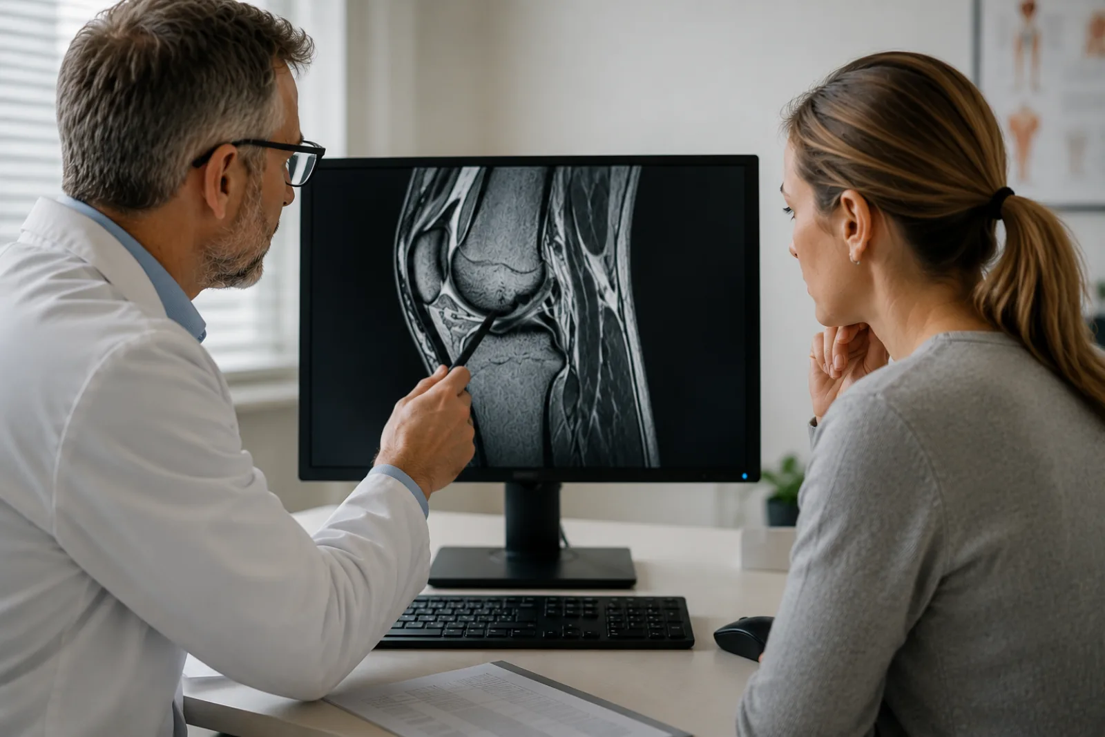
“Meniscal lesion, Stoller grade III.” Reading this sentence in a knee MRI report for the first time, most people understand only that something is not right — but not how serious it actually is. Next to it are terms like “signal alteration of the anterior cruciate ligament” or “grade II chondropathy,” each one precisely chosen. To the radiologist, this is everyday clinical language. To the patient, it is a text that ends up in a search engine late at night.
This article translates the most common terms in a knee MRI report into plain language — from the meniscus grading system to the signs of a cruciate ligament injury. It won’t replace the conversation with your doctor, but it should get you into that conversation knowing what the words mean.
What an abnormal knee MRI report does not automatically mean
An MRI of the knee is a sensitive scan. It shows structures that stay invisible on X-ray — menisci, ligaments, cartilage, bone marrow. That sensitivity has a downside: MRI also picks up changes that cause no symptoms and need no treatment.
A widely cited study from the Framingham cohort examined the knees of 991 people between 50 and 90 years of age on MRI — without any preselection for symptoms [1]. The result: 35 percent had meniscal changes. 61 percent of all meniscus tears in this study caused no symptoms — these people had had no knee pain at all in the previous month. Among men aged 50 to 59, meniscus tears were found in 32 percent of cases; among women of the same age, in 19 percent. From age 70 on, the rate exceeded 50 percent in both sexes.
A more recent study using high-resolution 3-Tesla technology found meniscus tears in 30 percent of completely symptom-free adults — including, for the first time, even bucket-handle tears, which until then had been considered clinically relevant in every case [3].
Not every finding that sounds alarming is a problem that needs to be treated. The crucial question is not what the MRI report says, but whether the finding matches the symptoms.
What the radiologist assesses on a knee MRI
Knee MRI reports tend to run long — even when almost everything in them is normal. That is because the radiologist describes every structure in the joint individually. A systematic report is not a catalog of problems; it is an inventory.
The structures the radiologist assesses, one after the other:
Menisci — the medial (inner) and lateral (outer) meniscus. Two crescent-shaped cartilage discs that sit as shock absorbers between the thighbone and the shinbone. The report describes their signal, their shape, and any tears.
Cruciate ligaments — the anterior cruciate ligament (ACL) and the posterior cruciate ligament (PCL). They stabilize the knee against uncontrolled forward and backward sliding. The report assesses their course, their fiber structure, and their signal.
Collateral ligaments — the medial and lateral collateral ligaments. Stabilizers on the sides of the knee. “Unremarkable appearance of the collateral ligament structures” simply means: the collateral ligaments look normal.
Articular cartilage — the gliding layer on the joint surfaces of the thighbone, shinbone, and kneecap. Cartilage damage is described in grades (chondropathy).
Bones — the thighbone (femur), the shinbone (tibia), the kneecap (patella). The radiologist looks for bone marrow changes, cysts, or fractures.
Soft tissues — joint capsule, synovial lining, muscle tendons, bursae, joint fluid. The radiologist works through these as well.
A long list is normal. What matters is the “Impression” section at the end — that is where the radiologist sums up what is clinically relevant.
A meniscus tear in the report — what grades I, II, and III mean
The Stoller classification: grades I to III
The Stoller classification describes how the meniscal tissue appears on MRI [2]. It is based on the question of whether a signal change reaches the surface of the meniscus or not.
Grade 0 — a normal meniscus. Uniformly dark on MRI (low signal), no abnormalities.
Grade I — a punctate signal increase inside the meniscus that does not extend to the surface. This is not a tear. It is an intrasubstance change, often age-related and of no clinical significance.
Grade II — a linear signal increase that also stops short of the surface. This is not a tear either. When surgeons went in and looked, 89 percent of menisci graded I or II turned out to be completely normal [2].
Grade III — the signal change reaches the articular surface of the meniscus. This corresponds to a tear. However: whether this tear causes symptoms and needs to be treated depends on the clinical situation, not on the grade alone.
“Stoller grade II signal increase in the posterior horn of the medial meniscus” — What this means in plain language: in the rear section of the inner meniscus, there is a change that does not extend to the surface. Not a tear, but the kind of wear that is normal at that age. No reason for treatment.
Tear patterns — what the terms in the report stand for
When a meniscus tear (grade III) is present, the report additionally describes the shape of the tear [4]. This information is relevant for treatment planning.
Horizontal tear — the meniscus splits in layers into an upper and a lower half. The most common pattern in degenerative tears, especially in patients over 40. Usually manageable without surgery.
Longitudinal tear (vertical tear) — a lengthwise tear running parallel to the rim of the meniscus. More often caused by injury than by wear. Often repairable with sutures, because it runs through the better-perfused zone.
Radial tear — perpendicular to the rim of the meniscus, running from the inside outward. It interrupts the hoop tension of the meniscus and impairs its function. Suture repair is often more difficult here.
Bucket-handle tear — a displaced longitudinal tear in which a meniscal fragment flips into the joint space. It can cause the knee to suddenly no longer straighten or bend fully. If the knee locks, see an orthopedist or go to an emergency department promptly.
Root tear — the meniscus tears away at its attachment point on the bone. Functionally, this is close to a total loss of the meniscus, because the hoop tension is completely abolished.
Medial vs. lateral meniscus — why location matters
The medial meniscus is affected far more often than the lateral meniscus. 98 percent of medial meniscus tears involve the posterior horn — the rear section. The lateral meniscus is more mobile, adapts better to joint movement, and tears less often from degeneration.
Blood supply plays a role in the chances of healing. The outer zone of the meniscus (near the base, the “red zone”) is better perfused and heals better. The closer the tear lies to the free inner rim (the “white zone”), the lower the chance of healing on its own.
A cruciate ligament tear in the report — partial tear, complete rupture, and everything in between
What the radiologist sees — primary and secondary signs
The report of a cruciate ligament injury rests on direct and indirect MRI signs [6].
Primary signs concern the ligament itself: the fibers are no longer continuous (discontinuity), the ligament shows an abnormal shape or an increased fluid signal. For complete tears, the sensitivity of MRI is 83 to 100 percent, the specificity 96 to 100 percent.
Secondary signs result from the mechanism of injury and involve other structures:
- Bone bruise (bone marrow edema) — contusion marks on the thighbone and on the posterior outer tibial plateau. This typical “pivot-shift pattern” arises when the knee buckles inward during the trauma. If both contusion marks are present, the probability of a cruciate ligament injury is over 90 percent.
- Anterior tibial subluxation — the shinbone sits too far forward relative to the thighbone. A displacement of 7 mm or more points to a complete rupture.
- PCL buckling — the posterior cruciate ligament kinks because the anterior one has lost its stabilizing function.
“Disrupted fiber structure of the ACL with diffuse signal increase on T2w” — What this means in plain language: the fibers of the anterior cruciate ligament are no longer continuous. This points to a complete tear.
“Bone bruise in the lateral femoral condyle and posterolateral tibial plateau” — What this means in plain language: the bone contusions in this typical location are an indirect sign of a cruciate ligament injury. They result from the mechanism of injury and usually heal on their own over weeks to months.
Partial rupture — the gray zone in the report
With partial tears, MRI reaches its limits. Sensitivity here is only 40 to 77 percent — meaning MRI cannot reliably distinguish a partial tear from an intact but swollen ligament [6]. That imaging findings and clinical reality do not always line up is not an error, but a well-known characteristic of radiological diagnostics.
“Signal alteration of the ACL with preserved continuity, DD partial rupture vs. mucoid degeneration” — What this means in plain language: the cruciate ligament shows a change, but the fibers are still continuous. Whether this is a partial tear or a harmless structural change cannot be determined with certainty by MRI alone. The hands-on exam by the orthopedic specialist (Lachman test, pivot-shift test) is what fills in the missing piece.
Not every signal change in the cruciate ligament is a tear — and not every tear needs surgery.
Other terms that frequently appear in a knee report
Beyond the meniscus and cruciate ligaments, the knee MRI report regularly describes other structures. Most of these accompanying findings describe reactions of the joint, not diseases in their own right.
Chondropathy grade I–IV (Outerbridge classification) — cartilage damage in four stages, from softening (grade I) to exposed bone (grade IV). Cartilage changes are common from about age 40 on and are often purely age-related. Not every chondropathy causes symptoms.
Bone bruise (bone marrow edema) — a contusion mark in the bone, visible as a bright spot in the otherwise dark bone marrow. It results from a blow or compression and usually heals on its own in six to twelve weeks.
Baker’s cyst (popliteal cyst) — a fluid-filled pouch at the back of the knee. Not a disease in its own right, but a consequence of irritation in the joint. The cyst itself is rarely treated — what matters more is finding out what is irritating the joint.
Joint effusion — excess fluid in the joint space. A nonspecific sign of irritation in the knee. The effusion itself says little about the cause — it can occur after an injury, with inflammation, or with wear.
Ganglion cyst — a small, gel-filled cyst, often at the meniscus or the ligaments. In most cases a harmless incidental finding.
Does every meniscus tear need surgery?
No. The distinction between a degenerative and a traumatic tear is essential for the treatment decision.
Degenerative tears develop gradually, mostly in patients over 40, frequently as horizontal tears. They are part of the normal aging process of the joint. The international ESSKA-AOSSM consensus of 2024 recommends a structured exercise program as the first measure: strengthening the leg muscles, training balance, stabilizing the hip and trunk — over at least twelve weeks, ideally guided by physical therapy [5]. The results of this approach are on par with surgical treatment. A Cochrane review confirms this as well: for degenerative meniscus tears, arthroscopic surgery is not superior to conservative treatment [7].
Traumatic tears result from acute injury, often during sports, frequently as longitudinal or bucket-handle tears. Here the indication for surgery is set more readily — especially with a locking knee, a combined cruciate ligament injury, or young, physically active patients. The trend is toward meniscal repair (preserving the tissue) rather than partial removal.
The factors that go into the decision: tear pattern, location, age, activity level, extent of symptoms, and accompanying injuries. It is not the report alone that decides whether to operate — it is how the knee troubles you in everyday life.
Much the same applies to the cruciate ligament. Not every cruciate ligament tear requires reconstruction. With minor instability and low athletic demands, a conservative path with targeted muscle strengthening can be considered — the trade-off is individual.
If surgery is recommended, a second opinion is a routine step, not a judgment on anyone. It is especially worth taking when no one raised the nonsurgical options with you.
What you can do when you are holding a knee MRI report
- Put the report in context — don’t google terms in isolation. Individual terms typed into a search engine often create more uncertainty than clarity. This article can help you understand the terminology — but it cannot replace the conversation with your doctor.
- Document your symptoms. When does the pain occur? Under what kind of strain? Since when? Is there locking, instability, or swelling? This is what lets your doctor put the imaging finding in context.
- Prepare for the appointment. An MRI report should be discussed in a conversation, not merely acknowledged. Possible questions: “What does this mean for me specifically?”, “Is treatment necessary — and if so, which?”, “Are there alternatives to surgery?”
- If surgery is recommended: get a second opinion. For certain elective procedures, this is a right enshrined in German law (Section 27b SGB V). Especially worthwhile when conservative options were not discussed or when the finding is ambiguous.
- Don’t decide under time pressure. With the vast majority of knee findings, there is no urgency. A considered decision beats a fast one. The only exception: if the knee locks acutely — then see an orthopedic specialist promptly.
Frequently asked questions
What does a knee MRI show?
MRI depicts soft-tissue structures particularly well — better than X-ray or CT. It shows the menisci, cruciate ligaments, collateral ligaments, articular cartilage, bones, and surrounding soft tissue. That is why it is the imaging test of choice when a meniscus or ligament injury is suspected. Bone fractures, on the other hand, are often detected faster and more reliably on X-ray or CT.
What does a grade 3 meniscus tear mean?
Stoller grade III means that the signal change on MRI reaches the articular surface of the meniscus. This corresponds to a tear. Whether this tear needs to be treated depends on the symptoms and the clinical examination — not on the grade alone. Many people over 40 have grade III findings without ever having had knee pain.
Can a meniscus tear heal on its own?
Small tears in the well-perfused outer zone (near the base, the “red zone”) can heal. Tears toward the free inner rim, or larger tears, generally do not heal on their own. But that does not mean they must be operated on — if they cause no symptoms, they can be managed conservatively.
What does a cruciate ligament tear look like on MRI?
In a complete tear, the fibers of the cruciate ligament are no longer continuous, and the ligament shows an increased fluid signal. Typical accompanying bone contusion marks (bone bruise) on the thighbone and shinbone are frequently found as well. With partial tears, the picture is less clear-cut — here, the clinical examination complements the imaging.
How long does it take to receive a knee MRI report?
The scan itself takes 20 to 30 minutes. The written report is usually available within a few days, sometimes on the same day. For specialized questions or external reading, it can take somewhat longer.
You have received a knee MRI report and would like an independent specialist assessment? At Second View, a board-certified radiologist reviews your report and your images — explained understandably, without time pressure.
This article explains general principles; it does not replace an in-person exam or advice from your own doctor. If the knee locks, gives way suddenly, or your symptoms flare up sharply, see a doctor or go to an emergency department right away. Last medical review: May 2026.
Sources
- Englund M, Guermazi A, Gale D et al. Incidental Meniscal Findings on Knee MRI in Middle-Aged and Elderly Persons. N Engl J Med 2008;359:1108-1115
- Stoller DW, Martin C, Crues JV 3rd et al. Meniscal tears: pathologic correlation with MR imaging. Radiology 1987;163:731-735
- Horga LM, Hirschmann AC, Henckel J et al. Prevalence of abnormal findings in 230 knees of asymptomatic adults using 3.0 T MRI. Skeletal Radiology 2020;49:1099-1107
- Nguyen JC, De Smet AA, Graf BK, Rosas HG. MR Imaging–based Diagnosis and Classification of Meniscal Tears. RadioGraphics 2014;34:981-999
- Prill R, Pujol N et al. The Formal EU-US Meniscus Rehabilitation 2024 Consensus (ESSKA-AOSSM-AASPT), Part II — Prevention, Non-Operative Treatment and Return to Sport. Knee Surg Sports Traumatol Arthrosc 2025
- Tiwari V et al. Magnetic Resonance Imaging of Anterior Cruciate Ligament Injury. StatPearls 2024
- Stable C et al. Arthroscopic surgery for degenerative knee disease. Cochrane Database Syst Rev 2022