Mammography: A Second Opinion on an Unclear Finding
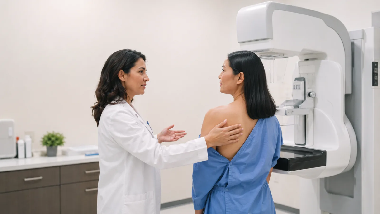
Four weeks after the screening, a second appointment is on the calendar. That was not the plan. The doctor’s office called — more testing was needed, the images had to be looked at again. Nothing more was said on the phone.
In the days between that call and the next appointment, the worry grows. Not because a diagnosis has been made — but because none has. Unclear, the report says, or abnormal, or a letter-and-number code like BI-RADS 0 that means nothing without medical training. And the mind keeps circling back to one question: does this mean cancer?
This article answers that question as honestly as it can. It explains what sits behind an unclear mammography finding, what the BI-RADS categories actually say, and why mammography sometimes cannot give a definitive answer. It shows what happens next — and when a radiological second opinion can sharpen the assessment. None of it replaces a conversation with your doctor; the point is to help you walk into that conversation knowing what to ask.
What an unclear mammography finding means — and what it does not
The word “unclear” sets off alarm bells. Most women read it as the first step toward a cancer diagnosis. In radiology it means something else: the images do not allow a reliable classification. Perhaps overlapping tissue is obscuring a structure, perhaps the tissue is too dense to judge, perhaps an abnormality cannot be confidently called benign.
“Abnormal” also sounds more threatening than it usually is. In radiology, it means only this at first: the radiologist saw something that should be looked at further. It does not mean that something is malignant. An unclear or abnormal finding is not a suspicion — it is a reason to look more closely.
The numbers from the German mammography screening program put this in perspective: of 1,000 women screened, about 30 are called back for further testing. Of those 30, 11 have a tissue sample taken. In 6, breast cancer is actually found [3]. So in 24 of the 30 women who get a recall, there is no cancer. The false-positive rate in the German screening program is 2.4 percent — well below the European limit of 5 percent [3].
A recall is not an alarm signal, then, but a built-in part of the screening program. It shows the process working carefully: better to look one time too many than to miss a change.
What the categories in your report mean — BI-RADS explained
Mammography findings are sorted using the BI-RADS system — the Breast Imaging Reporting and Data System of the American College of Radiology [2]. Every finding gets a category from 0 to 6, and each category comes with a concrete recommendation. BI-RADS is not a diagnosis; it is a recommendation for what to do next.
The categories at a glance:
BI-RADS 0 — Incomplete assessment. The images on hand are not enough for a reliable conclusion. More views, an ultrasound, or a comparison with earlier images are needed. This is the category behind most screening recalls — and the one that causes the most anxiety, even though all it means is that more information is needed.
BI-RADS 1 — Negative. The mammogram shows nothing out of the ordinary. Next screening in two years.
BI-RADS 2 — Definitely benign. There are findings to see, but they are clearly harmless — cysts, calcified fibroadenomas, or lymph nodes inside the breast. Again: back to regular screening in two years.
BI-RADS 3 — Probably benign. An abnormality that is very likely harmless. The probability of cancer is below 2 percent [2]. The recommendation is a short-interval follow-up after six months to document that it is stable. If nothing changes, the probability of malignancy drops further.
BI-RADS 4 — Suspicious. An abnormality that calls for a tissue sample. The category spans a wide range: BI-RADS 4a means a low probability of malignancy (2–10 percent), 4b an intermediate one (10–50 percent), 4c a high one (50–95 percent) [2]. The biopsy settles whether the abnormality is benign or malignant.
BI-RADS 5 — Highly suspicious. Strong suspicion of malignancy, with a cancer probability above 95 percent. A biopsy is urgently recommended.
BI-RADS 6 — Proven malignancy. A biopsy has already confirmed the cancer. At this point the mammogram serves treatment planning.
The categories that cause the most anxiety are BI-RADS 0, 3, and 4. All three say the same basic thing: there is an abnormality that needs a closer look. None of the three is a cancer diagnosis. And all three are built to name the next sensible step — not the worst case.
One common misunderstanding: many patients look up their BI-RADS category online and find probability figures for malignancy. Those numbers are statistical averages from large studies. They say nothing about any individual case. Your own finding only makes sense in conversation with the radiologist who read the images or with your doctor.
Why mammography findings can remain unclear
When a finding does not deliver a definitive answer right away, it rarely has anything to do with the quality of the reading. The most common reason for an unclear mammogram is dense breast tissue.
Breast density is graded on four levels in the ACR system [2]:
ACR a — mostly fatty tissue. Mammography passes through the tissue easily and changes stand out clearly. The best conditions for a reliable reading.
ACR b — scattered areas of dense glandular tissue. A few denser patches, but the breast is still straightforward to assess overall.
ACR c — heterogeneously dense tissue. Much of the breast is made up of dense glandular tissue. Smaller changes can be hidden.
ACR d — extremely dense tissue. This is where mammography runs hardest into its limits. Normal tissue and possible changes both show up white on the mammogram — it is like looking for a snowball in a snowstorm.
About 40 percent of women of screening age have breast density of ACR c or d. In them, the cancer detection rate drops measurably and the chance of a recall rises. This is not a flaw in the exam — it is a physical limit of the method.
Overlap adds to the problem. In conventional 2D mammography, three-dimensional tissue is projected onto a flat image, so structures lying one behind the other can stack up and mimic an abnormality — or hide one. The ToSyMa trial, a large multicenter study from Germany, showed that digital breast tomosynthesis (a form of 3D mammography) achieves a 48 percent higher detection rate in dense tissue than conventional mammography [5]. Not every facility offers it, but the data show where 2D mammography reaches its limits.
Radiologists in the screening program read hundreds of mammograms a week under standardized conditions. When one of them calls a finding unclear, that is not carelessness — it means the reader knows the limits of the method and would rather send an abnormality on for a closer look than let it slip through.
Since July 2024, mammography screening in Germany has covered women aged 50 to 75, which means roughly 2.5 million more women are being invited [6]. More scans also mean more recalls — and more women facing an unclear finding.
What happens after an unclear finding
The path from an unclear finding to an answer follows a set sequence. The screening program provides for the workup to happen within a few weeks [1, 3].
Additional imaging comes first. Which method is used depends on the finding:
Spot compression — targeted, magnified mammogram views of one area. They can show whether an apparent density is really there or was an artifact of overlapping tissue.
Breast ultrasound — especially helpful in dense tissue and for telling fluid-filled cysts from solid findings. Ultrasound uses no radiation and adds a second perspective to the mammogram.
Breast MRI — in certain cases the most sensitive method, especially with high breast density or when a finding cannot be pinned down on mammography and ultrasound.
The point of all this additional imaging is to see the abnormality from several angles and get a better sense of what it is.
A tissue sample (biopsy) is recommended when imaging alone cannot give a reliable all-clear — that is, with BI-RADS 4 and 5. The most common method is a core needle biopsy guided by ultrasound or mammography (stereotactic biopsy): minimally invasive, outpatient, under local anesthesia. It takes about 15 to 30 minutes. The histology result — the tissue diagnosis — usually comes back within a week and gives the definitive answer imaging could not.
The wait between the recall and the result is hard. That is not weakness; it is an understandable response to a situation that touches on existential questions. The fear in this phase is real and legitimate — and in the vast majority of cases it lifts once the result arrives. Until then, it helps to know how the workup runs and what the odds are: most recalls do not end in a cancer diagnosis.
When a second opinion is particularly worthwhile
Not every mammography finding needs a second opinion. But there are situations where a second look at the images can sharpen the assessment.
BI-RADS 3 vs. BI-RADS 4 — follow up or biopsy? The line between “probably benign, come back in six months” and “suspicious, biopsy recommended” has real clinical consequences. The decision turns on details — the shape, margins, and density of an abnormality — and different radiologists can weigh those details differently.
Conflicting assessments. When mammography and ultrasound point in different directions, or the radiologist who read the images sees it differently from the referring gynecologist, a second assessment can help resolve the discrepancy.
Dense breast tissue (ACR c or d). High breast density makes the reading objectively harder. A second look by an experienced radiologist can help place structures that are hard to make out in dense tissue.
Before deciding on a biopsy. When a tissue sample has been recommended and you are not sure it is really necessary, a second opinion can back up that recommendation or give a more nuanced sense of how urgent it is.
The data back this up: studies find discrepancy rates of 28 to 35 percent between first and second readings of mammograms [4, 7]. In a study at Memorial Sloan Kettering Cancer Center covering 790 patients, the second reading changed clinical management in 26 cases and turned up 10 additional cancers the first reading had not detected [4]. That does not mean the first reader was wrong — it shows how much judgment breast imaging involves. A second look is not distrust. It is due diligence.
What a radiological second opinion is — and what it is not
A radiological second opinion is an independent specialist assessment of the images you already have. The radiologist reviews the mammograms — plus any ultrasound or MRI images — and gives an independent evaluation.
What it is not: a clinical exam, a breast examination by hand, or new imaging. The second opinion works from the images that already exist. It does not replace the conversation with your doctor — it adds another specialist’s perspective to it. The second reader sees the same images as the first, but without the same time pressure and with one specific question to answer.
In practical terms: you can get a second opinion without booking another appointment for a physical exam. The images are sent digitally. The result usually comes back within a few days — a span that can make a real difference during an anxious wait. At best, the second opinion confirms the first assessment and puts your mind at rest. If it does not, it offers a more nuanced reading that can steer the rest of the workup more precisely.
Frequently asked questions
Can I take my mammography images with me? Yes. Patients have a right to their medical records, images included. Most doctors’ offices and screening units will give you the images on CD or as a digital dataset. A second opinion needs the original images in DICOM format, not just the written report.
What if the second opinion differs from the first? A difference is not a contradiction — it reflects how much interpretation breast imaging involves. The second reader may weigh details differently, categorize an abnormality differently, or spot additional findings. Take both assessments to your doctor, who can fit them into the overall picture.
Does a second opinion delay the workup? Usually not. A radiological second opinion based on digital images takes a few days — less than the usual wait between a recall and the workup appointment. You can request it in parallel with the regular process.
Is a second opinion worthwhile with BI-RADS 3? BI-RADS 3 is one of the categories where it can be most valuable. It sits right on the line between an all-clear and further testing, and whether an abnormality is called “probably benign” or sent for biopsy comes down to fine distinctions in the reading. A second look can confirm the category — or move the recommendation in another direction.
A radiological second opinion at Second View gives you an independent specialist assessment of your mammography images — one more perspective to bring to the conversation with your doctor.
Sources
- Leitlinienprogramm Onkologie (DKG, DKH, AWMF). S3-Leitlinie Früherkennung, Diagnostik, Therapie und Nachsorge des Mammakarzinoms, Version 5.0. AWMF-Register 032-045OL, 2025.
- American College of Radiology (ACR). BI-RADS — Breast Imaging Reporting and Data System, 5th Edition. ACR, 2013.
- Kooperationsgemeinschaft Mammographie. Evaluationsbericht Mammographie-Screening-Programm. mammo-programm.de, 2024.
- Coffey K et al. Second-Opinion Review of Breast Imaging at a Cancer Center: Is It Worthwhile? AJR Am J Roentgenol 2017.
- Heindel W et al. Digital breast tomosynthesis plus synthesised mammography versus digital screening mammography (TOSYMA): a multicentre, open-label, randomised controlled trial. Lancet Oncology 2022.
- DKFZ / Krebsinformationsdienst. Altersgrenze im Mammographie-Screening Programm auf 75 angehoben. 2024.
- Memorial Sloan Kettering Cancer Center. Second Opinion Following Routine Breast Imaging Supports Diagnostic Accuracy. 2019.
This article is general information and cannot replace an individual medical exam or consultation. If you notice changes in your breast — a lump you can feel, skin changes, a nipple that pulls inward, or discharge from one side — see your gynecologist promptly. Last medical review: May 11, 2026.