Understanding PI-RADS: What Your Prostate MRI Report Means
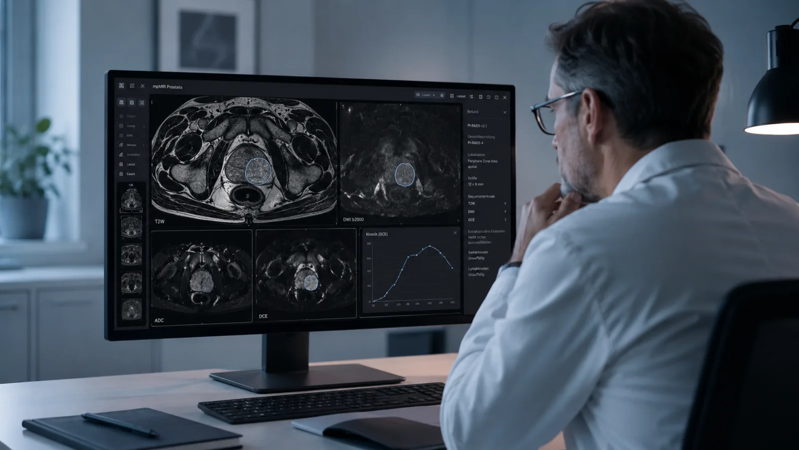
For most men, a prostate MRI report reads like a foreign language. Zones, sequences, terms with a Latin ring to them. And in the middle of it all sits a number: PI-RADS 4. It stands out because it looks nothing like the rest — short, clear, seemingly beyond argument. It feels like a verdict. Like a grade that has already been handed down.
That is exactly what it isn’t. A PI-RADS number is an estimate of probability — not a diagnosis. It tells you how suspicious the images look, not whether cancer is actually there.
This article explains what levels 1 through 5 mean, translates the terms you are most likely to find in your report, and describes what usually comes next. It can’t give you a personal diagnosis or a treatment recommendation — that belongs in the conversation with your doctor.
What PI-RADS actually is — a probability, not a diagnosis
PI-RADS stands for Prostate Imaging Reporting and Data System. It is an international standard that radiologists use to read magnetic resonance imaging (MRI) of the prostate the same way everywhere. The current version, 2.1, dates from 2019.
At its heart is a scale from 1 to 5 that describes how likely a clinically significant prostate cancer is. A 1 means very unlikely, a 5 very likely. The scale rates the images — not the tissue itself.
The phrase clinically significant is doing a lot of work. It means a cancer that should be treated because it can grow and spread. Not every prostate cancer belongs in that category. Some tumors grow so slowly that they never cause symptoms in a man’s lifetime. PI-RADS is aimed at the kind that can actually become dangerous.
Which leads to the most important point, right at the start: the number is a measure of probability. It tilts the assessment in one direction, but it proves nothing. Only a tissue sample can do that.
How MRI images become a number
To make sense of the number, it helps to know how it is arrived at. The scan is called multiparametric because it combines several types of images.
The T2-weighted sequence shows the tissue structure of the prostate in fine detail. Diffusion imaging — referred to in the report as DWI and ADC — measures how freely water molecules move through tissue. Densely packed tissue, of the kind tumor cells form, holds that movement back. The contrast-enhanced sequence (DCE) shows blood flow.
Which sequence carries the most weight depends on where the finding sits. The prostate has two important regions: the peripheral zone — the outer, larger portion, where most cancers start — and the transition zone around the urethra, which enlarges harmlessly with age. In the peripheral zone, diffusion imaging settles the question; in the transition zone, the T2-weighted sequence does.
In the report, that comes out looking something like this:
“T2-hypointense lesion in the peripheral zone with diffusion restriction.”
In plain language: at one spot in the outer part of the gland, the tissue looks darker than its surroundings on the T2-weighted images, and water movement is restricted on diffusion imaging. Together, those two findings make the spot look suspicious. The PI-RADS number boils observations like these down to a single rating.
What levels 1 through 5 mean
Each level can be tied to a rough probability that the finding hides a cancer that needs treatment. The figures below come from a pooled analysis of several studies compiled in the current guideline. One caveat first: these are reference values, not exact odds for any one man — they vary, sometimes widely, with the study, the group of patients, and the experience of the radiologist reading the scan.
- PI-RADS 1 — very unlikely: about 2 percent. No suspicious lesion.
- PI-RADS 2 — unlikely: about 4 percent. Probably benign.
- PI-RADS 3 — equivocal: about 20 percent. The gray zone (see the next section).
- PI-RADS 4 — likely: about 52 percent. A cancer that needs treatment is likely, but not proven.
- PI-RADS 5 — very likely: about 89 percent. High probability, often with signs of local spread.
Germany’s national prostate cancer guideline (version 8, 2025) ties clear recommendations to these levels. It is an S3 guideline — the highest evidence grade German medical societies award, reserved for recommendations built on a systematic review of the research. At PI-RADS 1 and 2, the guideline recommends against biopsy. At PI-RADS 4 and 5, it calls for targeted biopsy of the suspicious lesions, with two to three tissue samples each.
The number rarely stands alone in the report. A location usually follows:
“PI-RADS 4, index lesion in the left apical peripheral zone.”
In plain language: the spot that drives the rating — the index lesion — sits in the outer part of the gland on the left, near the tip of the prostate. PI-RADS 4 means “likely,” not “certain.” About half of these findings turn out not to be a cancer that needs treatment once the tissue sample is taken.
PI-RADS 3 — why the middle number causes the most uncertainty
Level 3 causes more uncertainty than any other. It is neither an all-clear nor a clear warning sign. About one in five PI-RADS 3 findings turns out to be a cancer that needs treatment — and four out of five do not.
PI-RADS 3 does not mean “a small cancer.” It means the images alone are not enough to decide. That is why the way this level is handled has changed recently.
Since its 2025 update, the S3 guideline advises against biopsying PI-RADS 3 findings right away when a man’s individual risk is low — and it gives that advice its strongest grade of recommendation. Whether the risk counts as low depends on other factors. The most important one alongside the PI-RADS level is PSA density.
PSA density sets the PSA value against the size of the prostate: the PSA value divided by the volume of the gland. The reasoning is simple. A large prostate that has enlarged harmlessly produces more PSA to begin with, so an elevated value there is less worrying than the same value in a small gland. The guideline uses about 0.15 ng/ml per milliliter of prostate volume as a point of reference. It is not a hard threshold above which a biopsy becomes necessary or below which it becomes pointless — it is one piece of the overall picture.
When the risk is low, a follow-up MRI is an alternative to taking tissue right away. If the finding stays the same over time, that argues against an aggressive process. If it changes, a targeted biopsy can follow.
Why two radiologists can arrive at different numbers
A PI-RADS number is a specialist’s judgment. It follows clear rules, but it is still a judgment — and judgments can differ. The studies show this plainly: the share of findings rated suspicious varies from center to center, as does the share later confirmed. Agreement between experienced readers is good, but not perfect — least of all at the border between PI-RADS 3 and 4.
That is not a sign of sloppy work. It is the nature of a hard call. Borderline cases look alike on the images. Image quality, technical settings, and how much experience the reader has with this particular question all feed into whether a lesion ends up a 3 or a 4. When radiological assessments diverge for structural reasons like these, that is normal variability — not a personal failing.
For you as a patient, that has a concrete consequence. At the border between 3 and 4, the rating is what decides whether a biopsy is recommended at all. An independent second reading of the same images can confirm the assessment — or shift it. Either way you gain clarity: a confirmation gives you confidence for the next step, and a correction can spare you a biopsy or send you to one that is properly targeted. There is more on how often radiological assessments disagree with one another elsewhere on this site.
Asking for that second read is not a vote of no confidence in the first report. It follows from what a probability estimate is — and it is an established part of careful diagnosis.
Other terms that frequently appear in the report
Beyond the number and the location, a few other terms turn up regularly. The most important ones:
- Index lesion: the lesion that determines the PI-RADS score. When several are described, it is usually the one with the highest level or the largest size.
- Peripheral zone and transition zone: the two main regions of the gland. Where a lesion sits influences how it is rated.
- PSA density: the PSA value relative to the volume of the gland.
One phrase worries men more than any other:
“Extracapsular extension cannot be excluded with certainty.”
In plain language: the images cannot say for certain whether the change reaches beyond the capsule of the organ. That is a question for planning treatment later — not a statement about whether the finding is benign or malignant. Uncertainty in a report is an honest answer, not a bad sign in disguise.
Finally, two things get confused often enough to be worth separating: PI-RADS and the Gleason score, which today is usually reported as the ISUP grade. They describe different things. PI-RADS estimates from the images how likely a cancer that needs treatment is. The Gleason or ISUP value describes how aggressive the tissue is — and it comes only from the biopsy. So the number in your MRI report says nothing about your Gleason score. It says how urgently the question needs answering.
What to do when you are holding a PI-RADS report
A PI-RADS report is a step along the way, not an endpoint. A few things help put it in perspective and prepare you for the next conversation.
- Read the number in context. The PI-RADS level only means something alongside the PSA value, the PSA density, and any earlier findings. A number on its own rarely tells the whole story.
- Come with specific questions. Good ones: Where is the index lesion — which zone, and how large? What is my PSA density? And why is a biopsy being recommended, or why not?
- With PI-RADS 3, keep asking. This is the level where your individual risk matters most: do the PSA density and the overall picture argue for a biopsy or for a follow-up MRI?
- Consider a second reading of the images. At the border between 3 and 4, or before a planned biopsy, an independent assessment of the images you already have can back up the decision — with no new scan needed.
One practical point about cost: in Germany, statutory health insurance has often not paid for a prostate MRI done before a biopsy, which left patients to cover it themselves as a private service. That is in flux now that the new guideline gives MRI a stronger role, so it is worth asking your insurer how your own case would be handled.
The situation resembles other unclear imaging findings — a lung nodule on CT, for instance, where the decision also comes down to weighing watchful waiting, follow-up, and intervention.
Frequently asked questions
Is PI-RADS 4 already cancer?
No. PI-RADS 4 means a cancer that needs treatment is likely — in studies, it is confirmed in about half of cases. The other half turn out to be benign changes. Only a tissue sample gives certainty.
At which level does a biopsy become necessary?
Under the current guideline, PI-RADS 4 and 5 get a targeted biopsy. At PI-RADS 1 and 2, the guideline recommends against one. PI-RADS 3 is the special case: when individual risk is low, no biopsy is done at first — instead the risk is weighed using factors such as PSA density.
What does PI-RADS 3 mean for me — biopsy or wait?
That depends on the whole picture, not the number alone. If your PSA density and your history point to low risk, a follow-up MRI is a recognized alternative to biopsying right away. Your doctor makes that call together with you.
Can a second reading change my PI-RADS number?
Yes — most often at the border between 3 and 4. Because the rating is a specialist’s judgment, an independent second read of the same images can land on a different level. It works from the images you already have; you do not need a repeat scan.
Does health insurance cover a prostate MRI?
In Germany, statutory health insurance has often not covered a prostate MRI done before a biopsy, which meant paying out of pocket. There are exceptions and case-by-case arrangements, and the picture is changing with the new guideline. Private insurers generally reimburse the cost.
A second opinion on your prostate MRI
At Second View, experienced board-certified radiologists read your existing MRI images independently and put the PI-RADS finding in perspective in a clear explanation written for patients. Before a decision about a biopsy in particular, a second specialist’s view of the images can give you the confidence to take the next step. A radiological second opinion is there to support the conversation with your doctor, not to replace it.
This article provides general information and cannot take the place of a personal medical exam or consultation. If you have symptoms or notice changes in your health, see a doctor. Last medical review: May 2026.
Sources and further reading
- Leitlinienprogramm Onkologie (Deutsche Krebsgesellschaft, Deutsche Krebshilfe, AWMF). S3-Leitlinie Prostatakarzinom, Version 8, 2025 (AWMF-Register 043-022OL; current version at www.leitlinienprogramm-onkologie.de).
- Turkbey B et al. Prostate Imaging Reporting and Data System Version 2.1: 2019 Update of PI-RADS Version 2. Eur Urol 2019.
- Oerther B et al. Cancer detection rates of the PI-RADSv2.1 assessment categories: systematic review and meta-analysis on lesion level and patient level. Prostate Cancer Prostatic Dis 2022.
- Westphalen AC et al. Variability of the Positive Predictive Value of PI-RADS for Prostate MRI across 26 Centers: Experience of the SAR Prostate Cancer Disease-focused Panel. Radiology 2020.
- Rosenkrantz AB et al. Interobserver Reproducibility of the PI-RADS Version 2 Lexicon: A Multicenter Study of Six Experienced Prostate Radiologists. Radiology 2016.
- Kasivisvanathan V et al. MRI-Targeted or Standard Biopsy for Prostate-Cancer Diagnosis (PRECISION). N Engl J Med 2018.