Runner's Knee: What Imaging Shows and When a Second Opinion Helps
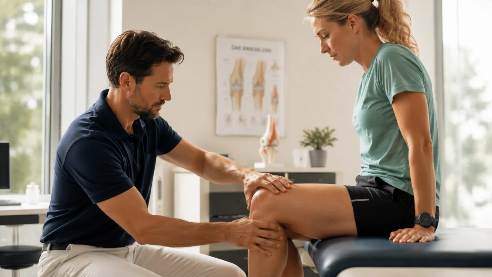
It is almost always the same point in the run. The first few kilometers feel fine, and then the knee starts to complain — a pulling sensation, sometimes a sharp pain that sharpens with every stride. Stop, walk for a few minutes, and you can feel it ease off. The next run, same story, same kilometer. That predictability is typical of what many people call “runner's knee.”
The term covers two different conditions, though, and there is a lot of confusion about which one anyone means. This article sorts them out and answers the question patients ask most often: do you need an MRI for this, and what does it mean if the scan turns something up? None of this replaces a conversation with your doctor — think of it as preparation for that conversation.
One name, two different knees — what “runner's knee” actually means
“Runner's knee” is not a formal medical term but an umbrella name. In German usage, it usually refers to iliotibial band syndrome (ITBS) — pain on the outer side of the knee. In English, “runner's knee” mostly means something else: patellofemoral pain, which sits at the front of the knee, on or behind the kneecap. An international expert panel even lists “runner's knee” as a synonym for that anterior knee pain [1].
So two people who both say “runner's knee” can be talking about two entirely different knees. Where the pain sits is the best guide.
If it hurts on the outer side of the knee, that points to iliotibial band syndrome. If the pain sits at the front, around the kneecap, that suggests patellofemoral pain.
A third problem often gets mixed in with these: jumper's knee (patellar tendinopathy), pain in the tendon just below the kneecap. It shows up mainly in jumping and stop-and-go sports, it is a separate condition, and it is not covered here. This article is about the two problems that typically come from running.
How runner's knee develops
Both forms are overuse injuries. A single misstep does not cause them; thousands of repetitions do — run after run, usually over weeks.
Iliotibial band syndrome centers on a tough band of tendon tissue: the iliotibial band, which runs down the outside of the thigh from the pelvis to just below the knee. Every time the knee bends and straightens, the band moves across a bony bump on the outer thigh, the lateral femoral epicondyle. The load peaks at about 30 degrees of flexion — the moment your foot hits the ground as you run. Friction and pressure then irritate the tissue underneath [2]. A rapid jump in weekly volume, a lot of downhill running, and weak hip-stabilizing muscles all make it more likely.
Patellofemoral pain is about the groove the kneecap slides in. If the kneecap does not track well in that groove, or takes heavy load over and over, the tissue responds with pain. Muscle imbalances at the hip and thigh are often part of the picture here too.
So runner's knee is less an injury than an overload reaction — the tissue is signaling that the current training load is more than it can absorb.
How to recognize runner's knee
The symptoms usually follow a recognizable pattern, and the pattern depends on which of the two conditions you have.
With iliotibial band syndrome, the pain sits in one spot on the outer side of the knee. It comes on with load, often at the same point in the run every time. Running downhill and climbing stairs typically make it worse, because they load the knee again and again at the critical flexion angle. At rest it usually settles quickly.
With patellofemoral pain, the picture is vaguer. Patients describe a dull ache at the front of the knee, around or behind the kneecap. It shows up on stairs, in squats, and running downhill — but also after sitting a long time with the knee bent, at the movies or in the car. That “moviegoer's sign” is a classic clue [1].
Where the pain sits and which movements set it off often say more about the cause than any image does. Those are the details your doctor will ask about first.
Do you even need imaging for runner's knee?
In most cases, no. Both forms of runner's knee are clinical diagnoses — a doctor makes them from your description of the symptoms and a hands-on exam. For patellofemoral pain, the international consensus statement says outright that imaging is rarely necessary [1]. The same goes for iliotibial band syndrome.
Imaging earns its place when it answers a specific question. That mainly happens in two situations: when another cause is suspected — meniscus damage, cartilage damage, a stress fracture — or when the symptoms will not settle despite consistent treatment. A scan then helps rule out conditions that can mimic runner's knee.
Each method has its own strength. An X-ray mostly shows bone and the position of the kneecap. Ultrasound looks at tendons and soft tissue, and can do so while the joint moves. MRI shows ligaments, cartilage, menisci, and soft tissue in the most detail, which is why it is the method of choice when a second condition or an alternative diagnosis needs to be investigated.
Runner's knee is diagnosed from how the symptoms behave and what the exam shows, not from an image. An MRI makes sense when a question is left over that the conversation and the exam cannot answer — not as the first step for every sore knee.
What MRI shows in runner's knee — and what an abnormal finding means
If you do end up having an MRI, the report can read a lot more alarming than the situation actually is. It helps to translate the terms.
With iliotibial band syndrome, the report often describes a signal change next to the outer thigh:
“Increased T2 signal in the soft tissue between the iliotibial band and the lateral femoral condyle.”
In plain language: there is fluid, or irritated tissue, between the tendon band on the outer side and the bone — what overload looks like on a scan [3].
With patellofemoral pain, it is usually the cartilage of the kneecap that turns up in the report:
“Grade II retropatellar chondropathy.”
In plain language: the cartilage behind the kneecap has started to roughen. Changes like this are common from middle age onward and, on their own, say very little about why you hurt.
Here is the crucial point: an abnormal finding is not the same thing as the cause of your pain. When researchers scanned pain-free adults with high-resolution 3-Tesla MRI, nearly every knee showed at least one abnormality [4]. And for iliotibial band syndrome, radiologists put it bluntly: you cannot make the diagnosis from the image alone — the finding means something only alongside the symptoms [3].
When images and symptoms fail to line up, that is not an oversight by the radiologist who read the scan. It is simply how radiology works.
When knee pain needs medical evaluation
Runner's knee is a nuisance, not an emergency. A few signs, though, suggest something else is going on and should be checked by a doctor promptly:
- The knee suddenly locks or gives way.
- The pain follows a clear fall or twisting injury, accompanied by rapid swelling.
- The pain occurs at rest or at night, unrelated to activity.
- The joint is red, warm to the touch, or accompanied by fever.
These point away from overuse and toward another cause — anything from a ligament injury to inflammation — that needs a workup of its own.
How runner's knee is treated — what surprises many people
The good news first: the vast majority of cases get better without surgery. The centerpiece is managing load — not necessarily a complete break from running, but cutting back for a while on volume, pace, and the things that trigger the pain, downhill running above all.
The second pillar is targeted exercise. For patellofemoral pain, the evidence for movement and strength training is the strongest: a Cochrane Collaboration review found consistently that exercise therapy reduces pain and improves function [5]. Strengthening the hips, core, and thighs, plus tweaks to running form, helps with iliotibial band syndrome as well.
Surgery is kept for rare exceptions — when symptoms drag on for many months despite consistent conservative treatment, for instance. With an overuse injury, patience almost always does more than an operation.
If surgery does come up, having a second specialist look at the images is a routine part of the process, not a vote of no confidence in your doctor — especially if the conservative options are not exhausted yet or the findings are ambiguous. As with the knee MRI report and the ankle, the same rule applies: what should drive the decision to operate is how much the symptoms cost you day to day, not the finding by itself.
What you can do if the pain keeps coming back
- Locate the pain instead of googling individual terms. Where does it sit, and which movement sets it off? Pain on the outside while running downhill points somewhere different from pain at the front on stairs. This article can get you oriented; it does not replace the conversation with your doctor.
- Keep track of your symptoms. How far into the run does the pain start? At what pace, on what terrain? How long has this been going on? Details like these are often worth more to the diagnosis than an image.
- Cut back your training early and get physical therapy. Reducing volume in time and doing targeted strength work usually shortens the whole episode considerably.
- Come to the appointment prepared. Good questions: which form of runner's knee is this? Is imaging necessary — and what question is it meant to answer? If you already have a written report, go through it together during the visit instead of just filing it away.
- If surgery is recommended, consider a second opinion. An independent read of the images you already have can confirm the decision without any new scan.
Frequently asked questions
Is “runner's knee” the same as patellofemoral pain syndrome?
Not necessarily. In German usage, “runner's knee” (Läuferknie) usually means iliotibial band syndrome, with pain on the outer side of the knee. In English, the same phrase more often means patellofemoral pain at the front of the kneecap. Where the pain sits tells you which one is meant.
Do I need an MRI for runner's knee?
Usually not. Both forms are diagnosed clinically, from your symptoms and a physical exam. An MRI is useful when another cause has to be ruled out — meniscus or cartilage damage, say — or when the symptoms will not settle despite treatment.
How long does it take for runner's knee to heal?
That depends on which form you have, how long it has been going on, and how much you train. Many overuse injuries improve within a few weeks with consistent load management and targeted exercise. The key is to act early instead of pushing on with the activity that set it off.
Does runner's knee require surgery?
Usually not. Treatment is conservative. Surgery is a possibility only in rare cases, when symptoms persist for a long time despite consistent therapy. Before taking that step, a second opinion is well worth the time.
Can I keep running with runner's knee?
Often you do not have to stop entirely — you have to get the dose right. Dialing back volume and pace for a while, steering clear of what triggers the pain, and adding strength work usually beats either quitting altogether or carrying on unchanged. Work out the specific plan with your doctor or physical therapist.
A second opinion on your knee report
At Second View, a board-certified radiologist reads your existing images independently and explains the findings in plain language. When it is unclear whether an abnormal finding really explains your pain — or when surgery is being scheduled — a second specialist view can give you the confidence to take the next step.
This article is for general information and cannot replace a personal exam or medical advice. See a doctor if the joint suddenly locks, if severe swelling follows a fall, or if you have pain at rest or at night or signs of inflammation. Last medical review: June 2026.
Sources
- Crossley KM, Stefanik JJ, Selfe J, Collins NJ et al. 2016 Patellofemoral pain consensus statement from the 4th International Patellofemoral Pain Research Retreat, Manchester. Part 1: Terminology, definitions, clinical examination, natural history, patellofemoral osteoarthritis and patient-reported outcome measures. Br J Sports Med 2016;50(14):839-843.
- van der Worp MP, van der Horst N, de Wijer A, Backx FJG, Nijhuis-van der Sanden MWG. Iliotibial Band Syndrome in Runners: A Systematic Review. Sports Med 2012;42(11):969-992.
- Flato R, Passanante GJ, Skalski MR, Patel DB, White EA, Matcuk GR Jr. The iliotibial tract: imaging, anatomy, injuries, and other pathology. Skeletal Radiol 2017;46(5):605-622.
- Horga LM, Hirschmann AC, Henckel J et al. Prevalence of abnormal findings in 230 knees of asymptomatic adults using 3.0 T MRI. Skeletal Radiol 2020;49(7):1099-1107.
- van der Heijden RA, Lankhorst NE, van Linschoten R, Bierma-Zeinstra SMA, van Middelkoop M. Exercise for treating patellofemoral pain syndrome. Cochrane Database Syst Rev 2015;(1):CD010387.